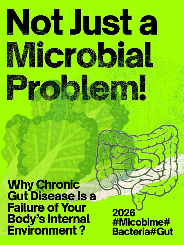
By Mohammed Attallah | Independent Systems Biology Researcher
You have been sick for a long time.
Not the kind of sick that shows up on a standard blood test. The kind that shows up at 2am when you are bloated, exhausted, and staring at the ceiling, calculating how many days it has been since you felt normal.
You tried the low FODMAP diet. Six weeks of strict elimination. You felt better. Then week seven arrived and the bloating returned. You tried rifaximin. Two weeks of improvement followed by the same symptoms at month three. You tried berberine and oregano oil. Your practitioner called the crash that followed “die-off.” You pushed through it. Nothing durably changed.
A stool test showed dysbiosis – the wrong bacteria in the wrong amounts. Practitioners treated the bacteria. The bacteria came back. Every single time.
Nobody asked the question that changes everything: why does the environment inside your gut keep recreating the conditions that allow these bacteria to thrive?
That is the question this work is built on. The answer is not in the bacteria. It is in the cells that line your colon and what has gone wrong with their energy production. The bacteria are not random villains. They are sophisticated organisms responding rationally to an environment that your own body is – unknowingly – creating for them.
I am not a physician and I do not have a formal science degree. What I have is years of deep investigation into the primary scientific research, driven by the need to understand why so many people fail every standard approach. I call the framework that emerged from that investigation the Host Capacity Model.
The Central Idea
The dominant model of chronic gut dysfunction treats it as a microbial problem. Too many bad bacteria, not enough good bacteria. Kill the bad ones, add the good ones, restrict the food they eat. This approach is not entirely wrong – but it is incomplete in a way that makes it fail predictably.
Bacteria do not exist in isolation. They live inside an ecological system – your gut – whose conditions are controlled by your own body. How much oxygen is in the gut lumen. How inflamed the lining is. What signaling molecules are circulating. What fuel sources are available. Whether the mucus layer is intact.
When your body maintains these conditions correctly, the gut environment is naturally hostile to pathogenic bacterial overgrowth. Good bacteria thrive, bad bacteria cannot establish dominance, and any dysbiosis that develops is temporary and self-correcting.
When these conditions fail – and specifically when the energy metabolism of the cells lining your colon breaks down – the gut shifts into a stable pathological state that is self-reinforcing and resistant to every intervention aimed at the bacteria themselves.
Treating the bacteria without fixing the environment is like trying to stop weeds from growing by pulling them out, while leaving the conditions that make the soil perfect for weeds completely unchanged. They grow back every time, not because your technique was wrong, but because the underlying problem was never addressed.
Part 1: The Oxygen Sink – The Master Switch That Controls Everything
Here is the fundamental biological fact that most gut health discussions never reach.
The cells lining your colon – called colonocytes – are responsible for maintaining the most important ecological condition in your gut: keeping it oxygen-free.
The entire colon is supposed to be anaerobic, meaning almost no oxygen. This is not a design flaw. It is the precise condition that allows the beneficial bacteria your gut depends on – organisms like Faecalibacterium prausnitzii and Roseburia intestinalis – to survive. These bacteria, called obligate anaerobes, die in the presence of oxygen. They absolutely require an oxygen-free environment. When oxygen enters the colon, they cannot survive, and the bacteria that replace them are exactly the ones associated with chronic disease.
So how does the colon stay oxygen-free? The colonocytes consume it.
Healthy colonocytes run primarily on a fuel called butyrate – a short-chain fatty acid that beneficial gut bacteria produce when they ferment dietary fiber. When colonocytes burn butyrate, they consume oxygen at the mucosal surface in the process. That oxygen consumption is what maintains the oxygen-free environment on the other side of the cell wall, in the gut lumen.
This is called the oxygen sink, and it is the master control switch of your entire gut ecology.
Research published in the journal Science confirmed this directly: when colonocytes are burning butyrate efficiently, the oxygen level in the gut lumen stays below 1% – the strict anaerobic conditions obligate beneficial bacteria need. When colonocyte butyrate oxidation is impaired for any reason, oxygen levels in the lumen rise, obligate anaerobes decline, and a family of bacteria called Proteobacteria – which includes Klebsiella pneumoniae, E. coli, and related organisms – expands to fill the space.
These Proteobacteria are facultative anaerobes, meaning they can survive in both oxygen-containing and oxygen-free environments. When the oxygen sink collapses and oxygen enters the lumen, they gain an ecological advantage over the bacteria that cannot tolerate it.
A 2024 study demonstrated this with direct measurement: reducing colonocyte butyrate oxidation alone – without any other intervention – was sufficient to increase luminal oxygen, decrease butyrate-producing bacteria, and increase Proteobacteria. The dysbiosis emerged directly and predictably from the colonocyte metabolic failure.
This is why dysbiosis is not, at its root, a microbial problem. It is a metabolic problem in the host tissue. The bacteria are responding to conditions you created. Fix the conditions, and the ecology fixes itself.
Part 2: The PPAR-γ Gate – How Inflammation Blocks the Repair Switch, and Why PPAR-γ Treatments Fail
The molecular switch that controls whether colonocytes burn butyrate or switch to a less efficient fuel is a protein called PPAR-γ (Peroxisome Proliferator-Activated Receptor gamma).
Think of PPAR-γ as a biological master switch inside the colonocyte. When butyrate enters the cell and activates PPAR-γ, this protein travels to the nucleus – the cell’s control center – and turns on the genes for efficient fat-burning metabolism. It switches on the machinery that burns butyrate, switches off the inflammatory signals that would otherwise generate nitrate in the gut lumen, and reinforces the cell’s barrier function. One switch activation produces multiple coordinated beneficial effects simultaneously.
When PPAR-γ is working, the colonocyte burns its fuel efficiently, the oxygen sink is maintained, inflammatory signaling is suppressed, and the barrier that keeps bacteria away from the immune surface is reinforced.
Here is the problem. When pathogenic gram-negative bacteria like Klebsiella shed a molecule called LPS (lipopolysaccharide) – a structural component of their outer membrane that gets released into the gut lumen – that LPS is detected by a sensor on colonocytes and immune cells called TLR4 (Toll-Like Receptor 4). TLR4 functions essentially as a bacterial alarm sensor. When it detects LPS, it fires off a cascade that activates a master inflammatory switch called NF-κB.
NF-κB travels to the nucleus and directly competes with PPAR-γ. They both need the same molecular machinery to do their jobs, and when NF-κB is active, it locks up that machinery for its own use, leaving PPAR-γ unable to function. NF-κB also triggers the production of inflammatory cytokines – specifically TNF-α and IL-1β – that actively reduce how much PPAR-γ protein the cell produces in the first place.
The result: PPAR-γ is simultaneously blocked from working and reduced in quantity.
This is why PPAR-γ agonist treatments – medications like rosiglitazone and pioglitazone, or natural compounds promoted as PPAR-γ activators – fail so reliably in patients with active gut inflammation.
Some practitioners prescribe these with sound logic: activate PPAR-γ, restore butyrate oxidation, close the oxygen sink. The target is correct. But you cannot meaningfully activate a receptor that is being actively blocked by a competing protein and simultaneously being reduced in the amount the cell produces. Applying a PPAR-γ agonist while NF-κB is fully active is like pressing a power button on a device whose circuit breaker has been tripped. The correct signal is being sent. The system that should respond to it is not functional.
The right sequence is: first substantially reduce NF-κB activity by addressing the LPS-TLR4 activation that is driving it; then allow PPAR-γ to recover; then support it with appropriate ligands. Applying the agonist before removing the blocker produces minimal effect at any dose.
PPAR-γ suppression also has a second critical consequence. One of the genes PPAR-γ normally keeps switched off is iNOS – inducible nitric oxide synthase, the enzyme that produces nitric oxide from the amino acid arginine. When PPAR-γ is suppressed and iNOS is released from that inhibition, the colonocyte begins producing nitric oxide. In the gut lumen, nitric oxide reacts with oxygen radicals generated by immune cells to produce nitrate (NO₃⁻).
Nitrate is the fuel that powers pathogenic bacteria’s most efficient respiratory system. The suppression of one cellular protein simultaneously collapses butyrate oxidation in the host and creates the high-energy fuel source that allows the pathogen’s survival machinery to run continuously. These two consequences spring from the same molecular event.
Part 3: The Bacteria – What They Are Running On, What It Costs Them, and Why They Cannot Be Killed
This is the most important section for understanding why antimicrobial strategies fail so reliably in chronic gut dysfunction. The bacteria in this situation are not passive targets waiting to be killed. They are running a sophisticated energy economy that your own inflammatory state is funding – and they are using that energy to maintain an array of survival mechanisms that make them essentially impervious to everything the standard clinical toolkit can throw at them.
The energy foundation: why nitrate changes everything
Every biological process – in bacteria as in human cells – requires energy in the form of ATP (adenosine triphosphate), the universal energy currency of cellular life.
When bacteria have access only to fermentation – breaking down sugars without using any electron-accepting molecule from the environment – they generate roughly 2–4 ATP per glucose molecule. This is energetically poor. The beneficial obligate anaerobic bacteria you want in your gut are limited to this pathway. They are metabolically austere and cannot afford expensive defense systems.
Nitrate respiration – using nitrate (NO₃⁻) as a fuel-completing molecule in place of oxygen – generates significantly more ATP than fermentation, giving Klebsiella and related bacteria a dramatic energy advantage over the bacteria they compete against. Research published in Science demonstrated that this host-derived nitrate gives these bacteria a measurable and significant growth advantage in the inflamed gut – an advantage that obligate anaerobes, which lack the genetic machinery for nitrate respiration, cannot share.
The bacteria use three separate genetic systems for nitrate respiration – encoded by gene clusters called narGHI, napAB, and narZYW. Having three redundant systems means this capability cannot be eliminated by targeting any single enzyme.
This energy surplus is not used for simple growth. It funds an extraordinary collection of survival programs that, running simultaneously, make direct bacterial killing strategies nearly impossible. Here is what that energy is buying, explained in plain terms.
Efflux pumps – the bacteria are pumping out everything you throw at them
The most important resistance mechanism Klebsiella carries is not an enzyme that destroys antibiotics – it is an active pump system called AcrAB-TolC.
This is a three-part molecular machine spanning both layers of the bacterial membrane. Think of it as a conveyor belt that continuously grabs unwanted molecules from inside the bacterium and ejects them directly into the environment outside both membranes. The three components – AcrA, AcrB, and TolC – work together as one connected system. AcrB is the pump inside the inner membrane. AcrA links it to TolC, which is a channel through the outer membrane. Together they create a continuous export pathway that the bacterium runs at high speed whenever it senses a threat.
The genes acrA, acrB, and tolC that encode this system are switched on by regulatory proteins – called MarA, SoxS, and RamA – that are themselves activated by the exact conditions of an inflamed gut: oxidative stress, bile acid exposure, and antibiotic challenge. The pump is most active precisely when you most want it inactive.
What does this pump export? Fluoroquinolone antibiotics like ciprofloxacin. Tetracyclines. Macrolides. Various beta-lactams. Bile acids. And – this is critical for the clinical gut health space – thymol, carvacrol, and cinnamaldehyde, which are the active antimicrobial components of oregano oil, thyme oil, and cinnamon. The bacteria do not passively tolerate these compounds. They actively eject them in real time. And they have the ATP from nitrate respiration to run this pump continuously without it compromising their other functions.
Without the nitrate energy supply, running this pump at high capacity would compete with growth and essential cellular maintenance. Restore colonocyte metabolism, close the oxygen sink, eliminate nitrate availability, and this cost becomes genuinely competitive with survival. This is the basis of the fitness cost strategy – not trying to kill the bacteria directly, but making their own defensive programs too expensive to sustain simultaneously.
Siderophores – stealing iron the host is trying to hide
When your immune system detects infection, it deliberately hides iron – an essential nutrient for bacterial growth – through a strategy called nutritional immunity. It increases production of hepcidin (a hormone that traps iron inside your cells), lactoferrin (a protein that grabs free iron in mucosal secretions), and lipocalin-2 (a protein that specifically seizes the bacteria’s primary iron-grabbing molecule before it can deliver iron to them).
Klebsiella responds by producing specialized iron-grabbing molecules called siderophores – small organic molecules with extraordinarily high affinity for iron, binding it tighter than most host iron-sequestering proteins.
Klebsiella carries four separate siderophore systems, each encoded by its own gene cluster:
Enterobactin, produced by the entABCDEF gene cluster, has one of the highest iron affinities of any small molecule ever measured. It costs roughly 6–8 ATP to make one molecule. However, lipocalin-2 can grab the enterobactin-iron complex before it delivers the iron to the bacterium – so the host has evolved a countermeasure.
Salmochelin, produced by the iroA gene cluster (iroBCDE genes), is essentially enterobactin with glucose molecules chemically attached to it. This modification – called glycosylation – prevents lipocalin-2 from recognizing it. It is a stealth siderophore that bypasses the host’s primary countermeasure. The bacterium has, through evolutionary time, developed a direct workaround to one of the host’s main defenses.
Yersiniabactin, produced by genes on what is called the high-pathogenicity island (irp1, irp2, fyuA), not only grabs iron but also sequesters copper and zinc, modulates immune responses, and contributes to biofilm formation. It costs approximately 8–10 ATP per molecule.
Aerobactin, often encoded on plasmids – small circular DNA elements separate from the main chromosome that can be shared between bacteria – works faster than enterobactin at delivering iron to the bacterium, making it useful for rapidly dividing populations.
Under the iron restriction that the host is deliberately imposing, the bacterium upregulates all four of these systems simultaneously, producing siderophores in massive quantities – each molecule a significant metabolic investment. It can sustain this only because nitrate respiration provides the energy surplus to fund it. Without that energy, iron restriction becomes genuinely lethal rather than just pressuring.
Capsule – physical armor around the bacterium
Klebsiella surrounds itself with a thick coat of polysaccharide molecules called the capsule. This coat is encoded by a region of the genome called the cps locus, containing 17–20 genes that collectively produce and assemble this protective layer. The capsule sits between the bacterium’s outer membrane and the environment, physically blocking complement proteins from punching holes in the membrane, preventing neutrophils and macrophages from engulfing the bacterium, deflecting antimicrobial peptides, and slowing the penetration of many antibiotic molecules.
The capsule is continuously shed and must be continuously rebuilt – requiring constant sugar synthesis and transport, all at ongoing ATP cost. Hypervirulent Klebsiella strains produce so much capsule that the organisms form thick strings when manipulated in the laboratory. The metabolic cost of this hypercapsule production is substantial, but the protection it provides is proportionally enormous.
Here is what makes this particularly difficult: when the immune system attacks, it signals the bacteria through their own environmental sensing systems (specifically a two-component signaling system called the Rcs phosphorelay) to produce more capsule in response. The quorum sensing network (discussed below) also drives increased capsule production when population density is high. The immune activation that is supposed to be killing the bacteria is simultaneously telling them to build thicker armor.
Heat shock proteins – maintaining function under stress
When bacteria face stress – elevated temperature from fever, oxidative damage from immune cells, antimicrobial compounds – they upregulate a family of proteins called heat shock proteins (HSPs). These are not passive bystanders. They are an active investment in survival.
DnaK (the bacterial version of human Hsp70) grabs proteins that are starting to unfold under stress and helps them fold back correctly, consuming one ATP per protein it helps. GroEL/GroES (the bacterial version of human Hsp60) functions like a protective barrel – it takes damaged proteins into its interior and refolds them, consuming up to 98 ATP per complete operating cycle. Lon protease identifies proteins too damaged to save and destroys them before they can cause problems inside the cell, consuming 1–4 ATP per peptide bond cleaved. ClpB disaggregates protein clumps that have already formed, extracting individual proteins for refolding – one of the most ATP-intensive processes in a stressed bacterial cell.
Under the conditions of an inflamed gut – mild fever, immune oxidative stress, antimicrobial treatment – all four of these systems run simultaneously. Their combined ATP cost, layered on top of siderophore synthesis, efflux pump operation, and capsule production, creates a genuine energetic crisis.
This is the core insight of what I call the fitness cost strategy: rather than trying to kill the bacteria directly – which they have evolved multiple overlapping defenses against – create conditions where their own survival programs become too expensive to sustain simultaneously. Make them choose between defenses instead of running all of them at full capacity on an unlimited energy budget.
Biofilm and dormancy – why the bacteria survive every treatment course and always come back
The bacteria in chronic gut dysfunction do not float freely in the gut lumen as individual cells. They live in structured communities called biofilms – organized populations embedded in a self-produced protective matrix of polysaccharides, proteins, and extracellular DNA, anchored to the mucosal surface.
The decision between forming a biofilm versus swimming freely is controlled by a molecule inside the cell called c-di-GMP. When c-di-GMP levels are high, the bacterium switches into biofilm mode – attaches, builds the matrix, reduces motility, and shifts to a stress-resistant behavioral state. When c-di-GMP is low, it disperses and resumes free-swimming behavior. In the inflamed gut, conditions consistently favor high c-di-GMP and stable biofilm maintenance.
Inside the biofilm, bacteria at different depths behave very differently. Surface cells are metabolically active – they produce the matrix, interact with the immune environment, and are what standard clinical tests detect. Cells in the deepest layers, closest to the gut wall, enter a state of near-complete dormancy – minimal metabolism, minimal protein production, minimal cell division.
These deep dormant cells are maintained by genetic systems called toxin-antitoxin (TA) systems. Here is how they work, explained plainly:
Inside each bacterium, two proteins are being continuously produced – a toxin and its antitoxin. The antitoxin is less stable than the toxin and must be continuously replenished. Under normal conditions, the antitoxin keeps the toxin neutralized and the cell functions normally. When stress hits – antibiotic pressure, nutrient shortage, immune attack – the antitoxin breaks down faster than it can be replaced. As antitoxin levels fall, the toxin accumulates and becomes active. The toxin then shuts down essential cellular processes, pushing the cell into a dormant waiting state.
The MazEF system: MazF is an enzyme that cuts messenger RNA – the molecular instructions cells use to produce proteins. When MazE (the antitoxin) is depleted and MazF activates, it cuts mRNA molecules throughout the cell, globally halting protein production. The bacterium enters dormancy where it is making almost nothing, requiring almost no energy, and presenting almost no targets for any drug to hit.
The RelBE system: RelE cuts mRNA at the ribosome – the cell’s protein-making machinery – during translation. RelB is its antitoxin. Under nutrient starvation, a global stress program shuts down metabolism broadly, with RelBE coordinating protein synthesis shutdown.
The HipAB system: HipA is an enzyme that adds a phosphate group to another key protein, halting protein production. HipA/HipB produces the highest proportion of dormant persister cells of any known toxin-antitoxin system. The phenotype is literally named “high persistence” because of how many cells in the population it drives into dormancy.
Persister cells are not antibiotic-resistant in the genetic sense – they carry no resistance mutations. They survive antibiotics because they have stopped doing everything that antibiotics target. Cell wall synthesis is halted (so beta-lactam antibiotics find nothing to block). Protein synthesis is stopped (so tetracyclines and macrolides find nothing to inhibit). DNA replication is paused (so fluoroquinolones find nothing to damage). The antibiotic finds nothing to kill.
When the antibiotic course ends, conditions in the lumen – oxygen still elevated, nitrate still available, immune activation still ongoing – trigger antitoxin production to resume, neutralizing the toxin, restarting protein synthesis, and returning the bacterium to active growth – into an environment that is still ecologically exactly what it was before treatment began.
This is not a treatment failure. It is the mathematically inevitable outcome of applying antimicrobial pressure to a population with a dormant subpopulation in an unchanged ecological environment. The only thing that prevents this relapse pattern is changing the ecological conditions so that the awakening population finds an environment it cannot exploit. That requires restoring colonocyte metabolism and the oxygen sink – not killing more bacteria more aggressively.
Quorum sensing – bacterial communication, and why inhibiting it backfires
Bacteria communicate with each other through chemical signals, coordinating collective behaviors based on how many of them are present. This is called quorum sensing – the bacterium is essentially counting its neighbors and deciding what behaviors are worth coordinating.
Klebsiella primarily uses a system called LuxS/AI-2. The enzyme LuxS produces a precursor that spontaneously forms autoinducer-2 (AI-2) – a universal signal molecule produced by hundreds of bacterial species. As population density rises, AI-2 accumulates. When AI-2 crosses a threshold concentration – signaling that the population is large enough for coordinated behavior to be worthwhile – it triggers a program that upregulates capsule production, activates adhesion structures for stronger mucosal attachment, matures the biofilm, synchronizes iron-grabbing molecule production, and initiates virulence factor release.
Many researchers and practitioners are now proposing quorum sensing inhibition (QSI) as a non-antibiotic strategy – disrupt the bacterial communication, prevent the coordinated virulence. The logic is sound on its surface.
The problem I have been investigating: the same AI-2 signaling system Klebsiella uses for virulence coordination is used by Faecalibacterium prausnitzii, Roseburia intestinalis, and Bifidobacterium species for their own community organization and coordination of butyrate production. Every compound that disrupts AI-2 signaling affects all AI-2-producing bacteria – the beneficial ones alongside the pathogenic ones.
Furthermore, certain QSI approaches that disrupt mature biofilms can trigger dispersal – releasing previously attached bacteria into a free-swimming state. In a patient with compromised gut barrier function, that dispersal event may be more clinically dangerous than the maintained biofilm, releasing bacteria into a state more capable of crossing the barrier.
The deeper issue is that QSI treats a communication problem rather than an ecological one. The bacteria communicate the way they do because they are in conditions that favor the behaviors their communication coordinates. Change the ecological conditions, and their communication will coordinate different, less dangerous behaviors without any pharmacological interference.
Part 4: Your Bacteria Are Running on Your Inflammation, Not Your Food
Research published in Science in 2013 demonstrated something that fundamentally changes the logic of dietary-based gut treatments: bacteria in the inflamed gut are not primarily running on dietary carbohydrates. They are running on nitrate generated by your own immune system as a byproduct of inflammation.
The inflammatory immune response produces nitric oxide through iNOS – the same enzyme that PPAR-γ normally suppresses. That nitric oxide reacts in the gut lumen to form nitrate. And Klebsiella and related Proteobacteria carry three redundant genetic systems – narGHI, napAB, and narZYW – specifically for using that nitrate as fuel, generating significantly more ATP from it than fermentative bacteria can generate from fiber breakdown.
A follow-up review confirmed why this matters so much ecologically: the ability to perform nitrate respiration is more highly conserved within the Enterobacteriaceae family than in any other group of gut bacteria. The reason Enterobacteriaceae dominate during inflammation is not random – it is because they are genetically equipped to exploit the specific fuel that inflammation generates, and obligate anaerobes are not.
Low FODMAP diets, elemental diets, and carbohydrate restriction reduce fermentable substrates for bacterial fermentation metabolism. They do not reduce host-derived nitrate. The dominant pathogenic bacteria have switched their primary fuel source to nitrate from inflammation. Dietary restriction addresses the fuel of the bacteria you want to grow – the obligate anaerobes that produce butyrate – while leaving the pathogenic population’s energy supply completely intact.
This is the mechanistic explanation for why these diets produce symptom relief without resolution. They make you more comfortable while leaving the underlying ecological conditions unchanged.
Part 5: The NAD⁺ Collapse – Why the Colonocyte’s Engine Cannot Restart
At the center of everything is a molecule called NAD⁺ – nicotinamide adenine dinucleotide. To understand why colonocyte metabolism fails and why it fails in a way that is so difficult to reverse, you need to understand what NAD⁺ actually does.
NAD⁺ performs two completely different but equally essential jobs in the colonocyte.
First, it is an electron carrier in the process of butyrate oxidation. When butyrate is broken down inside the mitochondria, electrons are released and transferred to NAD⁺, converting it to its reduced form NADH. NADH then delivers those electrons to the electron transport chain – the series of protein complexes that use them to generate ATP. Without enough NAD⁺ to accept electrons, the whole process backs up and butyrate oxidation slows down, regardless of whether the enzymes that perform it are present.
Second – and this is where things get more complex – NAD⁺ is the fuel that a family of regulatory proteins called sirtuins run on. Sirtuins regulate the activity of other proteins by removing chemical tags called acetyl groups from them, in a process called deacetylation. Critically, each deacetylation event consumes one molecule of NAD⁺ – it is used up, not recycled. When NAD⁺ falls, sirtuin activity falls directly with it.
What is acetylation and why does deacetylation matter?
Proteins inside cells are not simply on or off. They are regulated by chemical modifications that change their activity levels. One of the most important is acetylation – the attachment of an acetyl group (a small chemical tag) to specific amino acids in the protein. When a protein is acetylated, it is typically less active – think of acetylation as a lock placed on a molecular switch, or a cap placed over the working part of a protein. The protein exists, but its function is suppressed.
Deacetylation removes that lock and restores full activity. Sirtuins are the primary enzymes that perform this deacetylation, doing so in response to the cell’s energy status – specifically in response to NAD⁺ availability. When cells have adequate NAD⁺, sirtuins run continuously, keeping critical regulatory proteins in their active, unlocked states. When NAD⁺ falls, sirtuins slow or stop, and those proteins drift back into their locked, less active states.
SIRT1 – the protein that unlocks mitochondrial repair and controls inflammation
SIRT1 deacetylates (removes the lock from) a protein called PGC-1α, which is the master regulator of mitochondrial biogenesis – the process of building new functional mitochondria and maintaining the ones that exist. PGC-1α by default sits in an acetylated, locked, inactive state. When SIRT1 has adequate NAD⁺ and removes those acetyl groups, PGC-1α becomes fully active and travels to the nucleus, where it turns on the genes for building new mitochondria, assembling the electron transport chain components, and running butyrate oxidation. Without SIRT1 deacetylating PGC-1α, the cell cannot replace damaged mitochondria with new functional ones, cannot upregulate its electron transport chain, and its capacity for butyrate oxidation declines progressively even when butyrate is available – because the machinery to burn it is not being maintained.
SIRT1 also deacetylates a subunit of NF-κB called RelA/p65, at a specific site called lysine 310. Acetylated p65 drives maximal inflammatory gene transcription. When SIRT1 removes the acetyl group at K310, it reduces NF-κB’s inflammatory output – functioning as a molecular brake on the inflammatory cascade. When NAD⁺ is depleted and SIRT1 activity falls, this brake is released. NF-κB becomes progressively more active, driving more CD38 expression, more iNOS production, more inflammatory cytokines – in a self-escalating inflammatory state that the cell has lost its biological capacity to correct.
SIRT3 – the protein that keeps the mitochondria functional
SIRT3 works inside the mitochondria themselves. Its job is to keep the proteins of the electron transport chain in their active, deacetylated states. Its specific targets include Complex I of the electron transport chain (when SIRT3 keeps Complex I deacetylated, it runs efficiently; when SIRT3 fails and Complex I becomes acetylated, electron flow slows and electrons leak out as superoxide radicals instead of doing productive work), key enzymes of the TCA cycle (which must stay active for butyrate to be processed), and SOD2 – superoxide dismutase 2 – the primary antioxidant enzyme inside mitochondria. SOD2 converts superoxide radicals into harmless hydrogen peroxide. SIRT3 activates SOD2 by removing acetyl groups at specific sites. When SIRT3 fails, SOD2 becomes inactive, superoxide accumulates inside the mitochondria, and that superoxide damages the iron-sulfur clusters embedded in Complexes I, II, and III – the structural components that actually carry electrons through the chain. When these clusters are damaged, the electron transport chain loses structural integrity and function cascades further downward.
The three simultaneous drains that make NAD⁺ recovery nearly impossible without targeted intervention
NAD⁺ is not being depleted from one direction. It is being attacked from three directions simultaneously, while its production is being cut off.
CD38 – the enzyme that destroys NAD⁺: When LPS activates TLR4 and NF-κB, one of the genes NF-κB switches on is CD38. CD38 is an enzyme that sits on the surface of immune and epithelial cells and cuts NAD⁺ apart into smaller molecules the cell cannot use. It also destroys NMN – nicotinamide mononucleotide – which is one of the main precursor molecules the cell would use to remake NAD⁺. CD38 is attacking both the existing NAD⁺ pool and its replenishment supply at the same time. Research at the Mayo Clinic demonstrated that LPS administration directly increases CD38 activity in tissues and that this CD38 increase directly reduces NAD⁺ levels – confirming that every time your immune system responds to bacterial LPS, it drives CD38 to destroy your NAD⁺.
PARP enzymes – the DNA repair system that consumes NAD⁺ catastrophically: When DNA is damaged – which happens constantly in inflamed gut tissue from oxidative stress, bile acid exposure, bacterial metabolites, and inflammatory signaling – enzymes called PARPs (Poly-ADP-ribose polymerases) activate to repair the damage. They are the emergency repair crews. But they consume NAD⁺ at an enormous rate to fund their work. In an acute injury, this is manageable – the crisis passes, PARP shuts off, NAD⁺ recovers. In chronic gut inflammation, DNA damage is continuous rather than episodic. PARP runs permanently. The drain is permanent.
NAMPT suppression – the NAD⁺ production factory shut down: NAMPT (nicotinamide phosphoribosyltransferase) is the rate-limiting enzyme in the NAD⁺ salvage pathway – the primary route by which cells regenerate NAD⁺ from its breakdown products, recycling them back into functional NAD⁺. Inflammatory cytokines produced by NF-κB activation – specifically TNF-α and IL-1β – suppress NAMPT expression. The factory producing NAD⁺ is shut down at the same time CD38 and PARP are consuming the existing supply at maximum rate.
The complete failure loop: LPS from dysbiosis activates TLR4, TLR4 activates NF-κB, NF-κB upregulates CD38 (destroying NAD⁺) and activates PARP (consuming NAD⁺) and suppresses NAMPT (halting NAD⁺ production). NAD⁺ falls. SIRT1 loses its fuel and can no longer deacetylate PGC-1α – so mitochondria cannot be rebuilt – and can no longer deacetylate NF-κB – so inflammation escalates further and drives more CD38. SIRT3 loses its fuel and can no longer maintain electron transport chain function or activate SOD2 – superoxide accumulates, damages the structural components of the respiratory chain, mitochondrial function deteriorates further. Butyrate oxidation collapses. The oxygen sink fails. Oxygen and nitrate enter the lumen. Pathogenic bacteria bloom. More LPS. The loop starts again, tighter than before.
Every step in this chain is supported by direct published experimental evidence. This is not a theoretical model – it is a documented biological process that standard treatments fail to interrupt because they are applied downstream of where the failure originates.
Part 5B: The IAP Loop – The Hidden LPS Off-Switch That Butyrate Activates
There is a protective mechanism in the healthy gut that almost nobody in clinical gut health practice is discussing, despite being directly supported by strong research and being immediately relevant to why the LPS-inflammation cycle becomes unbreakable.
The mechanism involves an enzyme called IAP – Intestinal Alkaline Phosphatase – produced by colonocytes and secreted directly into the gut lumen. LPS triggers TLR4’s alarm response through a very specific part of its structure – phosphate groups attached to a region called the lipid A tail. IAP removes those phosphate groups through a process called dephosphorylation. Without those phosphate groups, LPS cannot bind TLR4 strongly enough to trigger the inflammatory cascade. The alarm signal is chemically deactivated in the gut lumen before it ever reaches the receptor that would set off inflammation.
Research published in PNAS showed that butyrate directly activates the IAP gene in colonocytes – when colonocytes are burning butyrate, they produce IAP, which is secreted into the lumen to deactivate the LPS that bacteria constantly shed. When colonocytes are burning butyrate efficiently, IAP production is high, LPS is being continuously deactivated, TLR4 activation is dampened, NF-κB stays lower, less CD38 is induced, NAD⁺ is preserved, PPAR-γ can function, butyrate oxidation is maintained. Butyrate burning produces the enzyme that deactivates the molecule that would otherwise shut down butyrate burning.
When butyrate oxidation fails, IAP production falls. The LPS deactivation system shuts down at the exact moment LPS production is highest – because the dysbiosis driving the metabolic failure is simultaneously flooding the lumen with bacterial endotoxin. The off-switch is disabled when the alarm is ringing loudest.
Research in severe COVID-19 patients confirmed this directly: patients with severe disease had significantly reduced IAP activity in their stool, explained by the depletion of butyrate-producing bacteria – demonstrating that this colonocyte-IAP-LPS circuit operates in exactly the way the framework predicts, in real human disease.
This adds a third reinforcing loop to the failure cascade: more dysbiosis produces more LPS, more LPS suppresses butyrate oxidation, reduced butyrate oxidation means less IAP, less IAP means more active LPS reaching TLR4, more TLR4 activation means more NF-κB and more CD38, which depletes more NAD⁺, which further suppresses butyrate oxidation. Three separate loops reinforcing each other simultaneously – which is why this pathological state is so extraordinarily stable and so resistant to any single intervention.
Part 6: Trained Immunity – Why Inflammation Continues Even After Bacteria Decrease
Research published in Science formally defined what is now called trained immunity: when innate immune cells – the monocytes and macrophages that form the front line of immune defense – are repeatedly exposed to LPS over months or years, they undergo a form of epigenetic reprogramming that makes them permanently more reactive.
Epigenetic reprogramming means changes to the way DNA is packaged, not changes to the DNA sequence itself. DNA in cells is wound around protein spools called histones. Whether the DNA wound around a histone is accessible for reading (and therefore whether the genes on it can be expressed) depends on chemical modifications to those histones. When innate immune cells are repeatedly exposed to LPS, specific activating marks – called H3K4me3 and H3K27ac – accumulate on the histones surrounding inflammatory gene promoters. These marks keep the chromatin in an open, accessible state. The inflammatory genes are kept in a perpetually ready-to-fire configuration.
The consequence: a small LPS signal that a naïve immune cell would respond to minimally now triggers a full-scale inflammatory response from a trained immune cell, because its inflammatory machinery is primed and ready. Even when bacterial load decreases, these epigenetically reprogrammed cells continue generating inflammatory signals at high level – because the training does not reverse when the stimulus decreases. The cell has learned a behavioral pattern that persists independently of the current bacterial load.
This is why patients who partially clear their dysbiosis still have elevated inflammatory markers, still have symptoms, still relapse. The bacteria have been partially cleared but the immune cells are still firing at the level they were trained to fire at. The inflammation is no longer primarily driven by bacteria – it is being maintained by immune cells that learned to be hyperreactive. Reversing this requires months of genuinely sustained reduced immune activation – the epigenetic marks are gradually overwritten only when the inflammatory stimulus is consistently reduced over time.
Part 6B: How Gut Inflammation Reaches Your Brain – The Tryptophan Pathway
When the gut immune environment is chronically inflamed, an enzyme called IDO (indoleamine 2,3-dioxygenase) is activated in gut cells and immune cells by the inflammatory cytokine IFN-γ. IDO intercepts tryptophan – an amino acid that normally serves multiple beneficial purposes – and redirects it into a pathway called the kynurenine pathway.
In a healthy gut, beneficial bacteria convert tryptophan into molecules called indoles – specifically indole-3-aldehyde and indole-3-propionic acid – that activate a receptor in colonocytes and immune cells called AhR (Aryl Hydrocarbon Receptor). AhR activation drives production of a cytokine called IL-22, which specifically supports epithelial barrier repair and mucus production. It also promotes immune tolerance – the calibrated non-reactivity that allows peaceful coexistence between the immune system and the gut’s microbial community. This is one of the primary molecular languages through which a healthy microbiome communicates with and maintains the gut lining.
When IDO is active and tryptophan is diverted to the kynurenine pathway, this signaling is lost. More importantly, the kynurenine pathway produces a compound called quinolinic acid, which activates NMDA receptors in the brain.
NMDA receptors (N-Methyl-D-Aspartate receptors) are the primary excitatory receptors for glutamate – the main stimulatory neurotransmitter in the brain – and they are essential for normal cognition and synaptic function. At normal activation levels, they are necessary for memory formation and cognitive function. When quinolinic acid from sustained IDO activation reaches the brain in excess and overstimulates these receptors, neurons are driven past their optimal activation level into excitotoxicity – they are overexcited, their energy demands exceed what they can supply, and the circuits governing working memory, emotional regulation, stress tolerance, and sensory filtering progressively deteriorate.
This is the molecular explanation for why people with chronic gut dysfunction experience brain fog, anxiety, cognitive difficulties, sensory sensitivity, and emotional dysregulation that does not respond to standard psychiatric treatment. These are not psychological responses to being chronically ill. They are neurological consequences of a specific biochemical signal – quinolinic acid from IDO activation – driven by the same inflammatory loop that is destroying the gut ecology.
Individuals with genetic variants that impair glutamate clearance – such as variants in the SLC1A1 gene that reduce astrocyte glutamate transporter expression, the proteins responsible for clearing excess glutamate from brain synapses – or variants in GRM3 affecting presynaptic glutamate braking, are significantly more vulnerable to neurological symptoms from this mechanism.
Part 7: Why Your Genetics Matter – And How to Look at Your Own
Not everyone with the same gut conditions develops the same severity of colonocyte metabolic failure. The reason is genetic – the specific variants each person carries in the genes governing every step of the failure cascade described in this article.
The following variants are clinically meaningful to this framework. They can be examined in 30x whole-genome sequencing (WGS) – which reads every position in your entire genome at 30-fold depth, detecting both common and rare variants – or through consumer platforms like 23andMe, which covers a selected set of common variants but misses many rarer functional ones. For the most complete picture, 30x WGS from services like Sequencing.com is significantly more comprehensive. The raw data from either source comes as a text file with an rsid identifier (a standardized reference number for each variant position), chromosome location, and your genotype for each position. You can search for any specific rsid using a basic spreadsheet search function, then look up the functional meaning of your genotype in published research databases like SNPedia.
TLR4 variants – how sensitive your cells are to LPS
The TLR4 gene encodes the LPS alarm sensor. Two variants – rs4986790 (which changes one amino acid in the part of TLR4 that contacts LPS, reducing its sensitivity) and rs4986791 (a second change in the same region) – reduce TLR4’s response to LPS stimulation. People who carry these variants, especially those carrying both, mount a weaker NF-κB response to the same LPS exposure. This means slower CD38 induction, slower NAD⁺ depletion, and more margin before colonocyte metabolic failure becomes established. Both variants are in 23andMe data and all WGS datasets.
SIRT1 variants – how efficiently you maintain the deacetylation brake
The variant rs12778366 sits in the promoter region of the SIRT1 gene – the DNA segment that controls how much SIRT1 protein is produced. The T allele is associated with higher SIRT1 production at baseline. More SIRT1 means more robust deacetylation of both PGC-1α (keeping mitochondrial biogenesis active) and NF-κB p65 (keeping the inflammatory brake functional), requiring greater NAD⁺ depletion before these regulatory mechanisms fail.
PPARGC1A variants – your mitochondrial repair capacity
The variant rs8192678 changes one amino acid in the PGC-1α protein itself – glycine at position 482 becomes serine. This Gly482Ser substitution reduces how effectively PGC-1α activates the mitochondrial biogenesis gene program under metabolic stress. People homozygous for the Ser482 allele (carrying two copies of it) show impaired mitochondrial recovery after damage – their colonocyte mitochondria rebuild more slowly after inflammatory injury, requiring longer sustained NAD⁺ restoration before oxidative capacity begins to recover. This is one of the most studied functional variants in the PGC-1α gene. It is covered by 23andMe and present in all WGS datasets.
CBS and SUOX variants – how much hydrogen sulfide stress your mitochondria face
The CBS gene (cystathionine beta-synthase) encodes an enzyme that produces hydrogen sulfide in the gut from the amino acids homocysteine and cysteine. At normal levels, H₂S is a signaling molecule. At high levels – in individuals with CBS overactivity variants, or in dysbiotic guts with high populations of H₂S-producing bacteria – H₂S directly inhibits Complex IV of the mitochondrial electron transport chain. Complex IV is the final step where electrons are transferred to oxygen. When H₂S blocks it, the entire electron transport chain backs up. Oxygen consumption drops. The oxygen sink begins to fail even before the NAD⁺-sirtuin cascade is fully activated, meaning two independent pathways are converging on the same collapse.
The SUOX gene (sulfite oxidase) converts sulfite – a toxic intermediate in H₂S metabolism – to safe sulfate for excretion. Reduced SUOX activity from functional variants allows sulfite to accumulate, adding additional mitochondrial toxicity. These variants are best detected in WGS.
CD38 variants – your baseline NAD⁺ vulnerability
Individuals vary in how much CD38 their cells produce at baseline, influenced by genetic variants including rs1130420 and rs3733769. Those with constitutively higher CD38 activity start with lower effective NAD⁺ reserves and deplete faster under equivalent inflammatory conditions – reaching the threshold of sirtuin failure at lower bacterial loads and lower LPS exposures than people with lower baseline CD38 activity.
MUC2 variants – the integrity of your bacterial separation layer
The MUC2 gene encodes the primary structural protein of the mucus layer in the colon – the gel-like barrier that physically separates bacteria from the epithelial surface. Variants that reduce MUC2 production or alter its structural properties thin this layer, increasing direct bacterial contact with the epithelial surface. More bacterial contact means more LPS reaching TLR4, more immune activation, stronger trained immunity induction, and amplification of every downstream step in the failure cascade. Rare functional MUC2 variants are best detected in WGS.
The important principle across all of these: no single variant determines outcome. The relevant question is how many risk variants you carry that all impair the same pathway step. Multiple variants converging on NAD⁺-sirtuin function – high CD38 expression, reduced SIRT1 activity, impaired SIRT3 function, reduced PGC-1α recovery – create a compounded vulnerability that is fundamentally different from carrying any one of them in isolation. The genetic picture is a system to be read as a whole, not a list of individual defects.
Part 8: Why Every Standard Treatment Fails – And Exactly Why
Low FODMAP and dietary restriction:
These strategies reduce fermentable substrates for bacterial fermentation metabolism. They reduce the gas production, osmotic load, and fermentation-driven symptoms that come from fermentation by all gut bacteria. But they do not reduce host-derived nitrate from iNOS activation. The dominant pathogenic bacteria are primarily running on inflammation-derived nitrate, not dietary carbohydrates. Removing carbohydrates addresses the fuel source of obligate anaerobes – the bacteria you want – while leaving the pathogenic bacteria’s primary fuel source completely intact. The result is genuine symptom improvement without ecological recovery.
Antimicrobial herbs:
The active antimicrobial compounds in oregano oil (thymol and carvacrol) and cinnamon (cinnamaldehyde) are substrates of the AcrAB-TolC efflux pump in Klebsiella and related pathogens. The bacteria actively pump them out in real time. Obligate anaerobes, which lack equivalent efflux capacity, are preferentially killed. When gram-negative bacteria are killed, their outer membranes release LPS into the lumen – spiking TLR4 activation, driving NF-κB, upregulating CD38, acutely depleting NAD⁺, and worsening the colonocyte metabolic state that created the dysbiosis in the first place. What practitioners call “die-off reaction” is frequently this exact mechanism – acute LPS toxicity from mass bacterial killing in a host whose IAP activity is already depleted and whose NAD⁺ reserves are already compromised.
Berberine specifically:
Berberine activates AMPK – a cellular energy-sensing protein that, when activated, is supposed to drive mitochondrial biogenesis through PGC-1α – by inhibiting Complex I of the mitochondrial electron transport chain. In a metabolically healthy colonocyte, this mild energetic stress triggers a beneficial adaptive response. In a colonocyte where Complex I is already dysfunctional from SIRT3 failure, where iron-sulfur clusters are damaged from superoxide accumulation, and where PGC-1α is acetylated and inactive because SIRT1 lacks NAD⁺, adding a Complex I inhibitor deepens the existing mitochondrial dysfunction. The AMPK signal fires correctly – but the program it is calling for cannot be executed because the infrastructure to execute it has been disabled. The signal is correct. The system to respond to it is not functional.
PPAR-γ agonists in active inflammation:
As established in Part 2: NF-κB in active inflammation suppresses PPAR-γ transcription (less receptor is produced) and sequesters the co-activators PPAR-γ needs to function (the receptor that does exist cannot work). Applying PPAR-γ agonists without first substantially reducing NF-κB activity produces minimal effect. The correct intervention target is PPAR-γ restoration. The sequence is wrong. Remove the inhibitory environment first, then apply the agonist.
Probiotics without ecological restoration:
Research published in Science stated directly that metabolic reprogramming of colonocytes to restore epithelial hypoxia represents a promising alternative to targeting microbes – because harnessing the host’s own ecological control mechanism is fundamentally more durable than introducing organisms into an unchanged hostile environment. A probiotic introduced into a gut where oxygen is elevated, nitrate is available, IAP is depleted, and trained immune cells are generating maximal cytokines faces conditions identical to those that eliminated the original beneficial community. Colonization is transient. The ecology re-establishes its pathological equilibrium when supplementation stops.
Bacterial dormancy – the guarantee of relapse:
Persister cells maintained by MazEF, RelBE, and HipAB toxin-antitoxin systems survive every antimicrobial course by becoming metabolically invisible to drugs. When the course ends and ecological conditions remain unchanged – oxygen still elevated, nitrate still available – they resume active growth and rebuild the population to original density. This is the mathematical inevitability of treating a population with a dormant subpopulation in an unchanged ecological environment. Only changing the ecological conditions prevents this outcome.
Part 9: The Connection to Colorectal Cancer
The colonocyte metabolic failure described throughout this article is mechanistically continuous with early colorectal carcinogenesis – and this connection is urgently underinvestigated.
Research has confirmed that the loss of butyrate oxidation and the shift of colonocytes toward glucose fermentation – called the Warburg shift – is an underlying feature of colon cancer, ulcerative colitis, and dysbiosis alike. The connection is not coincidental. It is mechanistic.
In early dysplastic cells that have undergone the Warburg shift – meaning their mitochondria are dysfunctional and they have switched to fermenting glucose – butyrate is no longer burned as fuel. It accumulates inside the cell instead. When butyrate accumulates and reaches the nucleus, it inhibits enzymes called HDACs (histone deacetylases). HDAC inhibition keeps the histone proteins around DNA in an acetylated, open state, which keeps genes for cellular differentiation and growth arrest switched on. This is the epigenetic brake – the mechanism by which butyrate tries to push early cancer cells back toward normal differentiated behavior and away from uncontrolled proliferation.
But this correction mechanism can only work if butyrate can reach the cancer cell. And in a gut that has been chronically dysbiotic, three failures prevent this simultaneously.
First, butyrate production is already depleted – the obligate anaerobe communities that produce butyrate have been diminished by the same ecological failure that preceded malignancy. Second, the MCT1 transporter that carries butyrate into colonocytes is downregulated by the exact same inflammatory cytokines (TNF-α and IFN-γ) that have been driving the chronic gut inflammation for months or years – so even the depleted butyrate that exists cannot efficiently enter cells. Third, the cancer cell’s own Warburg shift means it would not oxidize butyrate as fuel regardless – so butyrate does not even accumulate inside the cell at levels sufficient to inhibit HDACs, because the transporter is not delivering enough of it.
The epigenetic correction mechanism that might have caught early dysplastic cells and pushed them back toward normal behavior fails at every level simultaneously – production failure, transport failure, and metabolic failure converging on the same outcome.
The chronic gut dysfunction described in this article is not merely a statistical risk factor for colorectal cancer. It is a mechanistic disabling of the cellular defense system that represents the body’s primary line of protection against early malignant progression. This is a connection that current colorectal cancer prevention frameworks – which focus almost exclusively on post-hoc colonoscopy screening – do not address at the mechanistic level.
Part 10: The Logical Sequence That the Biology Requires
If you understand the mechanism, the intervention sequence becomes clear. Every step follows logically from the one before it.
Step 1 – Stop making the mitochondrial failure worse. Before introducing anything new, evaluate what is already being used. Berberine adds Complex I inhibition to cells already in Complex I failure. Antimicrobial herbs cause LPS spikes that drive CD38 and accelerate NAD⁺ depletion. Bile acids depolarize mitochondrial membranes. These interventions may have roles in appropriate contexts with correct sequencing – but applied to established colonocyte bioenergetic failure without addressing the upstream conditions, they compound the problem.
Step 2 – Restore NAD⁺. Without sufficient NAD⁺, SIRT1 cannot deacetylate and activate PGC-1α, and SIRT3 cannot maintain electron transport chain function. All downstream mitochondrial recovery depends on this. NMN and NR are the primary precursors that bypass the CD38-NAMPT bottleneck. NR has additionally been found to directly inhibit CD38 activity – simultaneously providing NAD⁺ precursor while reducing the enzyme most responsible for destroying it in the inflammatory gut context.
Step 3 – Reduce the LPS load driving CD38 upregulation. Not by killing bacteria (which releases more LPS from dying cell walls) but by reducing mucosal immune activation through conditions that limit direct bacterial-epithelial contact: mucus layer support, tight junction restoration, and allowing IAP activity to recover as butyrate oxidation improves.
Step 4 – Support PPAR-γ recovery after NF-κB activity genuinely falls. PPAR-γ agonist approaches – whether dietary or pharmacological – are only effective after NF-κB activity is substantially reduced. This is not about the dose of the agonist. It is about the sequencing relative to the inhibitory environment.
Step 5 – Reintroduce fiber after ecological conditions begin shifting. High fiber reintroduction before colonocyte oxidative capacity has recovered provides fermentation substrate that opportunistic bacteria exploit before obligate anaerobes can re-establish. The ecological restoration must follow the metabolic restoration, not precede it.
Step 6 – Monitor the markers that reflect actual mechanistic change. Calprotectin reflects mucosal inflammation. Urinary organic acid panels can detect mitochondrial dysfunction markers. Stool metagenomics tracks the ratio of obligate to facultative anaerobes – the key ecological metric. Symptom improvement alone does not confirm that the mechanism is actually changing.
This Is Not a Finished Model – It Is an Active Investigation
Every mechanism described in this article is supported by published primary research – the references are listed below and I encourage you to read the original papers.
But I want to be honest about what this framework is and is not.
It is mechanistically coherent – every causal step connects to experimental evidence and the model generates specific, testable predictions. It explains the failure patterns of standard treatments with molecular precision. It accounts for individual variation through a genetic framework that can be examined in real genomic data.
It is not a complete clinical protocol. There are places where I am drawing connections between established mechanisms in ways that have not yet been directly tested in chronic gut dysfunction specifically in human clinical trials. I have tried to identify those places honestly throughout rather than presenting hypothesis as established fact.
What I am building, article by article, is a complete mechanistic account of why chronic gut dysfunction persists, why standard treatments fail in the specific and predictable ways they do, and how the same upstream failure connects to conditions as serious as colorectal cancer.
The people who have failed every standard approach deserve a framework that takes their failure seriously – not as non-compliance, not as reinfection, not as bad luck, but as the expected biological outcome of treatments aimed at the wrong level of the problem.
That is what this work is for. And it is far from finished.
Mohammed Attallah is an independent systems biology researcher investigating gut ecology, colonocyte bioenergetics, the host-microbe interface, bacterial energy economics, and the mechanistic connection between chronic gut dysfunction and colorectal cancer risk.
References
1. Fueling the fire: colonocyte metabolism and its effect on the colonic epithelia. Critical Reviews in Food Science and Nutrition. 2025.
2. Litvak Y, Byndloss MX, Bäumler AJ. Colonocyte metabolism shapes the gut microbiota. Science. 2018;362(6418):eaat9076.
3. Byndloss MX, et al. Microbiota-activated PPAR-γ signaling inhibits dysbiotic Enterobacteriaceae expansion. Science. 2017;357(6351):570–575.
4. Park B, et al. Crosstalk between butyrate oxidation in colonocyte and butyrate-producing bacteria. iScience. 2024;27(9):110853.
5. Gonçalves P, et al. Butyrate and the fine-tuning of colonic homeostasis. International Journal of Molecular Sciences. 2021;22(6):3061.
6. Winter SE, et al. Host-derived nitrate boosts growth of E. coli in the inflamed gut. Science. 2013;339(6120):708–711.
7. Winter SE, Bäumler AJ. Dysbiosis in the inflamed intestine: Chance favors the prepared microbe. Gut Microbes. 2014;5(1):71–73.
8. Camacho-Pereira J, et al. CD38 dictates age-related NAD decline and mitochondrial dysfunction through an SIRT3-dependent mechanism. Cell Metabolism. 2016;23(6):1127–1139.
9. Chini CCS, et al. CD38 ecto-enzyme in immune cells is induced during aging and regulates NAD⁺ and NMN levels. Nature Metabolism. 2020;2(11):1284–1304.
10. Swamynathan MM, et al. Epithelial NAD⁺ depletion drives mitochondrial dysfunction and contributes to intestinal inflammation. Frontiers in Immunology. 2023.
11. Bai P, et al. PARP-1 inhibition increases mitochondrial metabolism through SIRT1 activation. Cell Metabolism. 2011;13(4):461–468.
12. Yeung F, et al. Modulation of NF-κB-dependent transcription and cell survival by the SIRT1 deacetylase. EMBO Journal. 2004;23(12):2369–2380.
13. Hirschey MD, et al. SIRT3 regulates mitochondrial fatty-acid oxidation by reversible enzyme deacetylation. Nature. 2010;464(7285):121–125.
14. Malo MS, et al. Intestinal alkaline phosphatase is a gut mucosal defense factor maintained by enteral nutrition. PNAS. 2008;105(10):3551–3556.
15. Lallès JP. Intestinal alkaline phosphatase: a review of its enzyme role in the intestinal barrier function. Microorganisms. 2022;10(4):746.
16. Araújo JR, et al. Intestinal alkaline phosphatase activity and efficiency are altered in severe COVID-19 patients. Gastro Hep Advances. 2023;2(7):911–917.
17. Hamarneh SR, et al. Intestinal alkaline phosphatase exerts anti-inflammatory effects against LPS by inducing autophagy. Scientific Reports. 2020.
18. Netea MG, et al. Trained immunity: a program of innate immune memory in health and disease. Science. 2016;352(6284):aaf1098.
19. Vuscan P, et al. Trained immunity: general and emerging concepts. Immunological Reviews. 2024.
20. Donohoe DR, et al. The Warburg effect dictates the mechanism of butyrate-mediated histone acetylation and cell proliferation. Cell Metabolism. 2012;16(4):466–477.
21. Zong W, et al. Disruption of intestinal oxygen balance in acute colitis alters the gut microbiome. Gut Microbes. 2024;16(1):2361493.
22. Lewis K. Persister cells. Annual Review of Microbiology. 2010;64:357–372.
23. Gerdes K, Maisonneuve E. Bacterial persistence and toxin-antitoxin loci. Annual Review of Microbiology. 2012;66:103–123.
24. Flemming HC, Wingender J. The biofilm matrix. Nature Reviews Microbiology. 2010;8(9):623–633.
25. Nikaido H. Multidrug resistance in bacteria. Annual Review of Biochemistry. 2009;78:119–146.
26. Russo TA, et al. Aerobactin mediates virulence and siderophore production under iron-limiting conditions. Infection and Immunity. 1999.
27. Hengge R. Principles of c-di-GMP signalling in bacteria. Nature Reviews Microbiology. 2009;7(4):263–273.